Pathophysiology of Radiation Caries
Head/neck radiotherapy (RT) damages serous salivary gland cells, causing severe hyposalivation with thicker, more acidic saliva. For example, the OraRad study found stimulated salivary flow fell to ~37% of baseline at 6 months (median) and only partially recovered to ~59% by 18 months. Low flow and reduced buffering capacity impair tooth self-cleaning. Cariogenic flora (Streptococcus mutans, Lactobacilli, Candida) increase dramatically, while decreased salivary immunoglobulins and antimicrobial peptides further reduce defense.
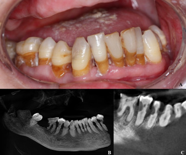
Radiation also directly weakens teeth. Microscopic studies show enamel/dentin become more brittle and demineralize more easily after RT. Odontoblastic damage and pulpal hypovascularity induce subsurface “radiation caries” and collagen breakdown. Clinically, lesions appear rapidly in non‑classic sites: circumferential cervical (“caries circularis”) and incisal/cuspal areas of incisors and molars. These lesions often progress without pain, and can destroy previously healthy teeth within months
Caries risk scales with dose: <30 Gy typically causes little tooth damage, whereas 30–60 Gy doubles to triples the risk and >60 Gy yields ~10× higher risk Importantly, RT that spares salivary glands markedly reduces this effect. In head/neck RT cohorts, parotid-sparing plans or tumor sites away from glands resulted in far fewer new caries. Thus, patients irradiated outside the head/neck (e.g. brain, non‑neck cancers) usually retain normal salivary function and do not develop the rampant “radiation caries” seen after head/neck RT.
Prevention Strategies
Proactive preventive care is critical. Fluoride therapy is universally recommended. High-fluoride regimens (e.g. 1.1% sodium fluoride gel daily in custom trays and 5% NaF varnish several times per year) are used to maintain enamel remineralization. Indeed, a recent prospective study found patients who used daily prescription fluoride had significantly less caries increment after RT. Many authors also add calcium-phosphate agents: e.g. casein phosphopeptide–amorphous calcium phosphate (CPP-ACP, e.g. MI Paste) twice daily along with fluoride. One report recommends 1.1% fluoride paste plus 2% CPP-ACP gel before/during RT to minimize demineralization. Note that weekly fluoride varnish and/or high-fluoride toothpaste (5000 ppm) are also advocated.
Saliva augmentation measures are used to compensate for xerostomia. Patients should avoid acidic beverages and add sugar-free chewing gum or candies (especially xylitol) to stimulate any residual flow. Pharmacologic sialagogues (pilocarpine or cevimeline) can be prescribed when some gland function remains, although effects vary. Humectants and saliva substitutes (viscous mucoadhesive gels) provide temporary relief of dry-mouth symptoms, though their caries-preventive efficacy is limited. Novel therapies like acupuncture or submandibular gland transfer (experimental) may be considered in research settings.
Oral hygiene and education are cornerstones. Patients should brush gently with a soft/ultrasoft toothbrush and fluoride toothpaste 2–3× daily, and floss or use interdental aids to minimize plaque Alcohol-free fluoride or bicarbonate mouth rinses are recommended (bicarbonate helps raise pH)Dentists must counsel patients and caregivers on the high caries risk. Dietary counseling to limit sugars (including sugary supplements or nutritional drinks) is crucial, as is quitting tobacco or alcohol which impair healing. Regular professional cleanings and fluoride applications (e.g. 3-month recalls) should be scheduled throughout RT and long-term. Multidisciplinary coordination (radiation oncologist, dentist, hygienist) ensures reminders and adherence.
Modern RT techniques should be leveraged. Intensity-modulated RT (IMRT) or volumetric arc therapy (VMAT) can spare at least part of the parotids/submandibular glands, reducing xerostomia and caries risk. Similarly, judicious RT dosing – keeping mean gland dose <26–30 Gy if possible – is advised. (By contrast, conventional 2D RT typically irradiates glands fully and nearly always causes xerostomia and caries.) NCCN/H&N oncology guidelines specifically advise custom fluoride trays and aggressive dental prophylaxis for all head/neck RT patients.
Clinical Management and Treatment
Pre-RT dental clearance: Before RT begins, patients undergo comprehensive dental exam. Unrestorable or severely diseased teeth are extracted with gentle technique and allowed ~10–14 days to heal before RTnature.com. All other teeth are restored or stabilized (deep caries filled, partial dentures adjusted, etc.)nature.com. Pre-RT prophylaxis (scaling, root planing) and topical fluoride are appliednature.com. Impressions are made for custom fluoride carriers. These measures reduce post-RT complications (including osteoradionecrosis).
Early detection: After RT, patients are followed closely. Surveillance visits are recommended at least every 3–6 months. At each recall, the dentist should inspect all tooth surfaces, use explorers/radiographs to find early lesions, and check for demineralization (often subtle at cervical margins). Fluorescence or digital imaging devices may aid detection of hidden lesions. Frequent monitoring allows intervention on incipient caries (e.g. resin infiltration, silver diamine fluoride, or sealants) before cavitation.
Restorative management: Dental restorations in the irradiated mouth pose challenges due to wetness, bonding issues, and high relapse risk. Glass-ionomer cements (GICs) or resin-modified GICs (RMGICs) are generally preferred. These materials bond via an acid-base reaction (less sensitive to moisture), release fluoride, and are relatively tolerant of dentin changes. In fact, one meta-analysis found markedly lower recurrent caries with GIC/RMGIC (0–26% at 2 yr) versus composite (26–44%) in irradiated patients. Newer high-viscosity GICs also help seal cervical lesions. Composite resins may be used for aesthetics when the field can be kept dry, but clinicians should expect higher failure rates. Amalgam is generally avoided (it can cause mucosal backscatter and local burns).
Any cavity preparations should be as conservative as possible. Sharp, overhanging edges from old restorations should be smoothed to prevent soft-tissue trauma. Secondary prevention (fluoride varnish, CPP-ACP pastes) should be applied regularly to restored teeth. Active incipient lesions may be managed nonrestoratively with high-concentration fluoride gels or silver diamine fluoride to arrest decay.
Endodontics vs extraction: The threshold for extraction is high after RT due to osteoradionecrosis (ORN) risk. Pulpally involved teeth are usually treated with root canal therapy if feasible. Endodontic treatment allows retention of the tooth and avoids bone injury. (Trismus or dry-mouth can make RCT difficult, but modified techniques – e.g. partial isolation, rubber-dam alternatives – can be used.) “Hopeless” teeth – roots fractured, or with severe periodontal disease – still may require removal, but only after preventive measures (e.g. antibiotics, hyperbaric oxygen protocols in select cases) and with surgical precautions (atraumatic extraction, primary closure).
Prosthetic rehabilitation: Removable prostheses (dentures) should have a hygienic design (supragingival borders) and fit precisely to avoid sores. Dentures should be checked/adjusted before RT to avoid trauma, and patients are often advised to avoid wearing them during acute mucositis. Fixed crowns/bridges are usually postponed or avoided unless absolutely needed, since margin leakage is likely. Simple overdentures or implants may be considered long-term, but implant placement in irradiated bone has higher failure (84% survival vs ~95% non-irradiated) and remains controversial.
Long-term follow-up: This population needs lifelong maintenance. Dental visits every 3–6 months should continue indefinitely, with ongoing fluoride applications and hygiene reinforcement. Patients must be counseled that any new caries should be addressed immediately, as lesions progress extremely quickly. Periodic adjuncts (chlorhexidine rinse for mutans control, motivational interviewing, reminder calls) are often used. In all, an “aggressive” preventive regimen must be maintained: intensive hygiene instructions, daily fluoride/CPP-ACP use, diet control, and prompt repair of any defects.
In summary, radiation caries arises from RT-induced xerostomia and tissue changes that create a highly cariogenic environment. Its prevention requires multidisciplinary vigilance: gland-sparing RT planning, patient education, and aggressive topical therapy (fluoride, remineralizers). When caries does occur, early detection and fluoride-boosted glass-ionomer restorations help salvage teeth in these challenging patients. With proper preventive care and follow-up, the severe dental decline once characteristic of head/neck RT can be mitigated.
Sources: Peer-reviewed studies and guidelines (Cancer Med 2017; Natl J Maxillofac Surg 2015; J Dent Res 2023; Br Dent J 2023; etc.) were used to compile this review

found that useful? let us know what you think